Painting 3D engineered muscle
Description of the photo
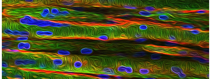
3D engineered muscle construct morphology was analyzed by IF: Sarcomere A-Actin immunofluorescence visualized by confocal fluorescent microscopy (in green) confirmed the alignment of the myoblasts and myotubes. Dystrophin (in red) satins mature myotubes. Nuclei were stained with DAPI (in blue)
Author
Name: Orfi Zakaria
Affiliation: CHU Sainte-Justine
Edition: 2020, Winner of the 3rd prize of the competition.


